This site is for healthcare professionals only. Please click here if you are not a healthcare professional.
 UK (English)
 Español
 Türkçe
 Deutsch
 Italiano
 Русский
 Français
Forget your password?
Clinical evaluations

Clinical evaluations
EEG with low-frequency intermittent photic stimulation and other clinical tests can be used to detect CLN2 disease1,2
The majority of children with CLN2 disease have been reported to show a response to low-frequency intermittent photic stimulation (IPS).3
EEG with IPS
EEG with low-frequency IPS can be used to detect photosensitivity in children with CLN2 disease
- A photoparoxysmal response (PPR) is common in patients with CLN2 disease and has features that are distinguishable from PPRs seen in other epilepsies3
- Low-frequency (1-2 Hz) IPS induces the appearance of time-locked high-amplitude occipital spikes in many cases3,4
- Positive PPR results should be followed by enzymatic and/or molecular testing to rule out NCL4
- Not all children with CLN2 disease will exhibit this response.3,4 Performing diagnostic enzymatic and/or molecular tests will rule out or confirm CLN2 disease if suspected4

EEG used with permission from Nicola Specchio, MD, PhD.
Many children with CLN2 disease show a photoparoxysmal response or a pathognomonic response to EEG with low-frequency (1-2 Hz) IPS.3
Other clinical assessments may provide findings indicative of CLN2 disease, including:
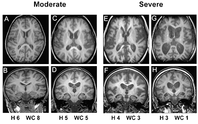
Brain MRI
Early in the disease, a brain MRI may appear normal or show subtle abnormalities. Abnormalities that should raise the level of suspicion for CLN2 disease include:1,2
- Cerebral atrophy, which is a common finding in NCLs
- Periventricular white matter hyperintensity
Later in the disease, cerebral atrophy becomes evident and will progress.1,2
Worgall S et al. Neurology. 2007 Aug 7;69(6):521-35.
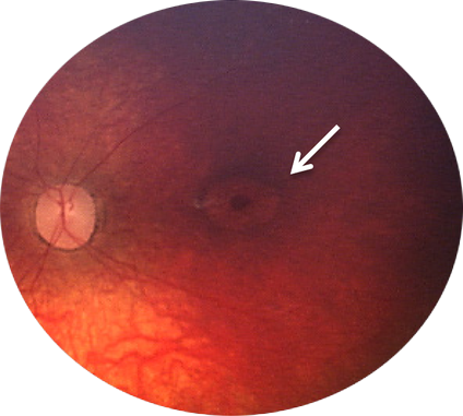
Ophthalmological assessment
Though blindness occurs late in the progression of CLN2 disease, visual abnormalities may be identified using certain ophthalmological assessments.5
Note: This image is from a 8-year-old patient, reflecting late-stage advances of CLN2 disease.6
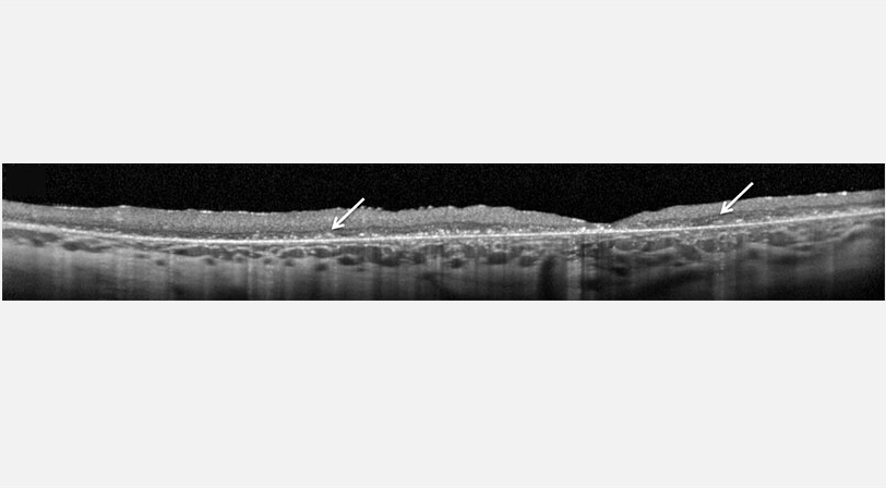
Ocular coherence tomography (OCT)
OCT can demonstrate progression of CLN2 disease via retinal degeneration and accumulation of hyper-reflective material.6,7
Fluorescein angiography (FA)
FA can be used to evaluate vascular leakage.6
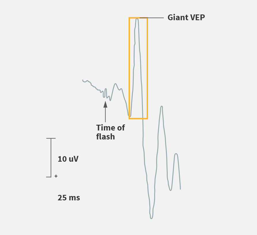
Visual evoked potential (VEP)
VEPs are abnormally enhanced until late in the disease and diminish in the final stage of the disease.1
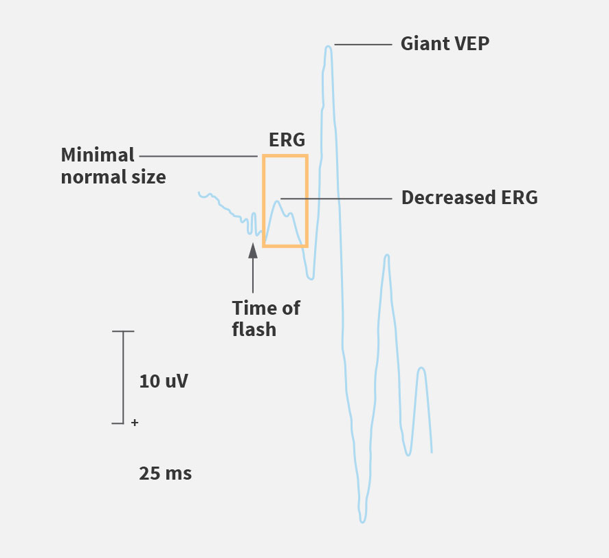
Electroretinogram (ERG)
ERG may be diminished before visual deterioration is clinically detected.1,2
Additional clinical tests used to identify CLN2 disease
Electron microscopy (EM)
Although the use of electron microscopy in clinical practice has decreased worldwide, it is used to detect the distinct curvilinear structure of ceroid lipofuscin in suspected cases, often when other tests are inconclusive.5
There are a number of clinical tests that can help identify CLN2, but only enzymatic and/or molecular testing can definitively diagnose CLN2 disease.
References: 1. Chang M et al. CLN2. In: Mole S, Williams R, and Goebel H, eds. The neuronal ceroid lipofuscinoses (Batten Disease). 2nd ed. Oxford, United Kingdom: Oxford University Press; 2011:80-109. 2. Mole SE et al. Correlations between genotype, ultrastructural morphology and clinical phenotype in the neuronal ceroid lipofuscinoses. Neurogenetics. 2005;6:107-126. 3. Albert DV et al. Unique Characteristics of the photoparoxysmal response in patients with neuronal ceroid lipofuscinosis type 2: can EEG be a biomarker? J Child Neurol. 2016;31:1475-1482. 4. Fietz M et al. Diagnosis of neuronal ceroid lipofuscinosis type 2 (CLN2 disease): Expert recommendations for early detection and laboratory diagnosis. Mol Genet Metab. 2016; 119:160-167 5. Mole SE and Williams RE. Neuronal ceroid-lipofuscinoses. 2001 Oct 10 [Updated 2013 Aug 1]. In: Pagon RA, Adam MP, Ardinger HH, et al., editors. GeneReviews® [internet]. Seattle, WA: University of Washington; 1993-2016. 6. Orlin A et al. Spectrum of ocular manifestations in CLN2-associated Batten (Jansky-Bielschowsky) Disease correlate with advancing age and deteriorating neurological function. PLoS One. 2013;8:e73128. 7. Williams RE et al. Management Strategies for CLN2 Disease. Pediatr Neurol 2017;69:102–112.